Alargamientos Óseos

Alargamientos Óseos
Dr Alejandro Robles Mendoza

viernes, 27 de marzo de 2015

Fijación de fracturas de la diáfisis femoral de longitud estable en los niños más pesados: clavos flexibles vs clavos rígidos bloqueados / Fixation of length-stable femoral shaft fractures in heavier children: flexible nails vs rigid locked nails

Fuente
Este artículo es originalmente publicado en:
http://www.ncbi.nlm.nih.gov/pubmed/21150726
http://journals.lww.com/pedorthopaedics/pages/articleviewer.aspx?year=2011&issue=01000&article=00005&type=abstract
De:
Garner MR1Bhat SBKhujanazarov IFlynn JMSpiegel D.
 2011 Jan-Feb;31(1):11-6. doi: 10.1097/BPO.0b013e31820321ab.
Todos los derechos reservados para:
Copyright © 2015 Wolters Kluwer Health, Inc. All rights reserved.

Abstract

BACKGROUND:

The treatment of femoral shaft fractures in heavier children has been studied extensively, however, no study has directly compared elastic nail (TEN) and rigid locking nails (RLN) in this population. Our goal was to compare TEN with RLN in length-stable diaphyseal femur fracturesin heavier children and adolescents (47 to 85 kg) using weight-matched cohorts.

METHODS:

We retrospectively reviewed records for length-stable diaphyseal femoral fractures treated with TEN or RLN over 8 years at our Level 1 Pediatric Trauma Center. Perioperative and follow-up data, including total charges for care, were recorded and radiographic images were reviewed. These data were used to create 2 cohorts having each patient in the TEN cohort matched to within 2 kg of the corresponding patient in the RLN cohort.

RESULTS:

Fifteen patients from each cohort could be weight matched (TEN, 60.8 kg vs. RLN, 60.4 kg). The RNL cohort was older (15.4 vs. 13.5 y; P = 0.005). Time in operating room and estimated blood loss were greater in the RLN cohort: 158 versus 220 minutes (P = 0.003) and 42 versus 182 ml (P = 0.003), respectively. All patients had a full range of motion at the latest follow-up. Complications were observed in 6 of 15 TEN and 10 of 15 RNL (P = 0.14). Implant-related problems were more common in RLN patients, but this was not statistically significant (3 of 15 vs. 9 of 15; P = 0.06). In the TEN cohort, malunion and leg length discrepancy (> 2 cm) each occurred in a single patient (20 degrees varus, 2.3 cm shortening, respectively) compared with 0 of 15 in RLN (P = 0.48). Treatment with TEN resulted in a total charge of $742 more than RLN (P = 0.75).

CONCLUSIONS:

In our weight-matched comparison, the use of TEN resulted in decreased time in operating room, estimated blood loss, and implant-related problems. Malunion and leg length discrepancy remain of concern when heavier patients are treated by TEN, but were not significantly increased relative to RNL in this study.

Resumen
ANTECEDENTES:
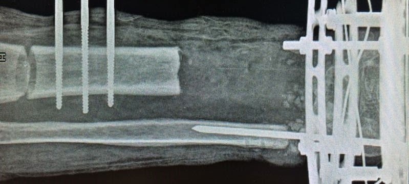
El tratamiento de las fracturas de la diáfisis femoral en niños más pesados ​​ha sido ampliamente estudiado,  sin embargo, ningún estudio ha comparado directamente los clavos elásticos (TEN) y los clavos de bloqueo rígido (NLR) en esta población. Nuestro objetivo fue comparar TEN con NLR en fracturas de longitud estable diafisaria del fémur en niños más pesados ​​y adolescentes (47 a 85 kg) utilizando cohortes emparejado con el peso.

RESULTADOS:
Quince pacientes de cada cohorte podrían peso igualados (TEN, 60,8 kg vs NLR, 60,4 kg). La cohorte RNL era mayor (15,4 vs. 13,5 y; P = 0,005). Tiempo en la sala de operaciones y la pérdida de sangre estimada fue mayor en la cohorte del NLR: 158 frente a 220 minutos (p = 0,003) y 42 frente a 182 ml (p = 0,003), respectivamente. Todos los pacientes tenían un rango completo de movimiento en el último seguimiento. Se observaron complicaciones en 6 de 15 TEN y 10 de 15 RNL (P = 0,14). Problemas relacionados con el implante fueron más frecuentes en los pacientes del NLR, pero esto no fue estadísticamente significativa (3 de 15 frente a 9 de 15; p = 0,06). En los diez cohortes, consolidación viciosa y la pierna longitud discrepancia (> 2 cm) cada ocurrieron en un solo paciente (20 grados de varo, 2,3 cm de acortamiento, respectivamente) en comparación con 0 de 15 en NLR (P = 0,48). El tratamiento con TEN resultó en un cargo total de $ 742 más que el NLR (P = 0,75).
CONCLUSIONES:
En nuestra comparación emparejados por peso, el uso de los TEN resultó en menor tiempo en la sala de operaciones, la pérdida estimada de sangre, y los problemas relacionados con el implante. Mala unión y la longitud de la pierna discrepancia siguen siendo motivo de preocupación cuando los pacientes más pesados ​​son tratados con TEN, pero no aumentaron significativamente con respecto a RNL en este estudio
PMID:
 
21150726
 
[PubMed - indexed for MEDLINE]

martes, 24 de marzo de 2015

Pérdida ósea segmentaria en las fracturas pediatricas de la extremidad inferior: indicaciones y resultados del transporte óseo / Segmental bone loss in pediatric lower extremity fractures: indications and results of bone transport

Fuente
Este artículo es originalmente publicado en:
http://www.ncbi.nlm.nih.gov/pubmed/25665166
http://journals.lww.com/pedorthopaedics/pages/articleviewer.aspx?year=2015&issue=03000&article=00005&type=abstract
De:
Arslan H1Özkul EGem MAlemdar CŞahin İKişin B.
 2015 Mar;35(2):e8-e12. doi: 10.1097/BPO.0000000000000392.
Todos los derechos reservados para:
Copyright © 2015 Wolters Kluwer Health, Inc. All rights reserved.


Abstract

BACKGROUND:

In this study, we evaluated the results of external bone transport, which was applied to 11 patients with traumatic bone loss who had not completed their bone development.
METHODS:

The average age of the 9 male and 2 female patients was 10.6 (range, 8 to 16) years. Eight of the defects were located in the tibia, whereas the other 3 were in the femur. The average defect was 5.4 (range, 4.5 to 8.5) cm. External bone transport was applied in the early period in 7 patients, whereas in 4 patients it was performed due to nonunion. Bifocal osteosynthesis and single osteotomy were performed in 2 patients with type B2 nonunion. Compression to the nonunion region and lengthening in the osteotomy region were applied. In 2 patients with type B1 nonunion, and the other 9 patients who had external bone transport, the gap was eliminated by bifocal osteosynthesis, single osteotomy, and bone transport to the osteotomy line.
RESULTS:

The mean follow-up period was 21 (range, 13 to 48) months. Complete union was achieved in all patients without any bone operation or graft application. No refracture was observed after the removal of the external fixator, and the average hospitalization time was 16 (range, 7 to 65) days. The average external fixation time was 4.2 (range, 3.5 to 5.5) months, and the mean external fixator index was 0.8 months (23 d/cm). The mean bone healing time was 5.1 (range, 4.6 to 6) months.
CONCLUSIONS:

To initially consider the open fractures with true or in situ bone loss in children as "anticipated nonunion," and determine the treatment strategies regarding this fact, may prevent nonunion and shorten the healing period. Bone transport in the treatment of traumatic bone defects in children is an easy biological procedure, with lower complications but higher success ratios.
LEVEL OF EVIDENCE:

Level IV-therapeutic.

Resumen

ANTECEDENTES:
En este estudio se evaluaron los resultados del transporte óseo externo, que se aplicó a 11 pacientes con pérdida ósea traumática que no habían completado su desarrollo óseo.
MÉTODOS:
La edad media de los pacientes 9 hombres y 2 mujeres fue de 10,6 (rango, 8-16) años. Ocho de los defectos estaban localizados en la tibia, mientras que el otro 3 estaban en el fémur. El defecto promedio fue de 5,4 (rango, 4,5 a 8,5) cm. Transporte óseo externo se aplicó en el período temprano en 7 pacientes, mientras que en 4 pacientes se realizó debido a la falta de unión. Osteosíntesis Bifocal y solo osteotomía se realizaron en 2 pacientes con pseudoartrosis tipo B2. Compresión a la región seudoartrosis y el alargamiento en la región de osteotomía se aplicaron. En 2 pacientes con pseudoartrosis tipo B1 y los otros 9 pacientes que tenían transporte óseo externo, la brecha fue eliminado por osteosíntesis bifocales, solo osteotomía, y transporte óseo a la línea de osteotomía.
RESULTADOS:
El período medio de seguimiento fue de 21 (rango, 13-48) meses. Unión completa se logró en todos los pacientes sin ninguna operación de médula o de la aplicación del injerto. Sin nueva fractura se observó después de la eliminación del fijador externo, y la estancia media fue de 16 (rango, 7-65) días. El tiempo medio de fijación externa fue de 4,2 (rango, 3,5 a 5,5) meses, y el índice de fijador externo media fue de 0,8 meses (23 d / cm). El tiempo medio de curación del hueso era 5,1 (rango, 4,6 a 6) meses.
CONCLUSIONES:
Para considerar inicialmente las fracturas abiertas con cierta o en la pérdida de hueso situ en los niños como "falta de unión anticipada", y determinar las estrategias de tratamiento con respecto a este hecho, se puede impedir la seudoartrosis y acortar el periodo de cicatrización. Transporte óseo en el tratamiento de defectos óseos traumáticos en los niños es un procedimiento biológico fácil, con las complicaciones más bajas, pero porcentajes de éxito superiores.

Nivel de evidencia:
Nivel IV-terapéutico

PMID:
 
25665166
 
[PubMed - in process]


lunes, 23 de marzo de 2015

Anatomy Of The Adductor Magnus Muscle

Publicado el 09/03/2015
Educational video describing the anatomy of the adductor magnus muscle.

Este vídeo es publicado originalmente en:

lunes, 16 de marzo de 2015

La innovación es importante, pero también imparte nuevo nivel de riesgo, complicaciones potenciales / Innovation is important, but also imparts new level of risk, potential complications

Fuente
Este artículo es originalmente publicado en:
http://www.healio.com/orthopedics/business-of-orthopedics/news/print/orthopedics-today/%7B0d5a4f05-92f4-489d-97e5-6efa11ccb482%7D/innovation-is-important-but-also-imparts-new-level-of-risk-potential-complications

De:
Anthony A. Romeo, MD; M. Michael Khair, MD
Todos los derechos reservados para:


©2015 All Rights Reserved.


Comparison of First- and Second-Generation Fixed-Bearing Total Ankle Arthroplasty Using a Modular Intramedullary Tibial Component

Fuente
Este artículo es originalmente publicado en:
http://www.ncbi.nlm.nih.gov/pubmed/25769492
http://fai.sagepub.com/content/early/2015/03/13/1071100715576568.abstract
De:
Lewis JS Jr1Green CL2Adams SB Jr3Easley ME3DeOrio JK3Nunley JA3.
 2015 Mar 13. pii: 1071100715576568. [Epub ahead of print]
Todos los derechos reservados para:
© The Author(s) 2015.
Copyright © 2015 by American Orthopaedic Foot & Ankle Society


Abstract

BACKGROUND:
This series reviews the clinical and radiographic outcomes of patients who underwent total ankle replacement (TAR) using first- and second-generations of a modern fixed-bearing prosthesis utilizing a modular intramedullary stem.
METHODS:
A consecutive series of first- and second-generation primary TARs with modular intramedullary stems were identified. Clinical outcome data were collected prospectively-including visual analog scale for pain, American Orthopaedic Foot and Ankle Surgery hindfoot-ankle, Short Musculoskeletal Function Assessment, and Short Form-36 scores. Preoperative coronal plane deformity and correction of deformity after TAR were assessed. Complications, subsequent procedures, and failure rates were compared. A total of 193 first- and 56 second-generation patients were identified with a mean follow-up of 3.7 and 2.1 years, respectively.
RESULTS:
Clinical outcome data reflected significant improvements at 1 year postoperatively, and improvements were maintained at 2-year follow-up for each group. Improvement in visual analog scale scores were significantly better in the second-generation group at 1 year postoperatively, but this was not maintained at 2 years. Mean coronal tibiotalar angles for ankles with preoperative varus or valgus deformities were significantly improved. Correction was maintained until final follow-up, with no significant differences in deformity improvement between groups. The rate of reoperation at 2 years postoperatively on the affected foot or ankle subsequent to the index ankle replacement for patients in the first-generation group (18.5%) was higher compared to the second-generation group (15.9%), but the time until reoperation was not statistically significant (P = .376). The implant failure rate was higher in the first-generation group (6.0%) compared to the second-generation group (2.6%) at 2 years postoperatively, but the time until failure was not significantly different (P = .295).
CONCLUSION:
Patients who underwent TAR with a first- or second-generation fixed-bearing prosthesis with an intramedullary tibial component demonstrated significant improvements in all measures of pain and function with sustained improvements in coronal plane alignment. The second-generation prosthesis demonstrated slightly better improvements at 1 year and was associated with lower reoperation and implant failure rates.
LEVEL OF EVIDENCE:

Level II, comparative series.

© The Author(s) 2015.

KEYWORDS:

INBONE; ankle arthritis; ankle arthroplasty; total ankle replacement


Abstracto

ANTECEDENTES:

Esta serie se revisan los resultados clínicos y radiológicos de los pacientes sometidos a reemplazo total de tobillo (TAR) con primer y segundo generaciones de una moderna prótesis de soporte fijo que utiliza un vástago intramedular modular.

MÉTODOS:

Tallos se identificaron una serie consecutiva de alquitranes primarios de primera y de segunda generación con intramedular modular. Datos de los resultados clínicos se recogieron de forma prospectiva, incluyendo una escala analógica visual para el dolor, American Orthopaedic Foot Cirugía de Tobillo y posterior del pie-tobillo, corto Evaluación de la función musculoesquelética, y Short Form-36. Preoperatoria deformidad plano coronal y la corrección de la deformidad después TAR fueron evaluados. Las complicaciones, los procedimientos posteriores, y las tasas de fracaso fueron comparados. Un total de 193 de primera y 56 pacientes de segunda generación se identifica con un seguimiento medio de 3,7 y 2,1 años, respectivamente.

RESULTADOS:

Datos de los resultados clínicos reflejan mejoras significativas a 1 año después de la operación, y las mejoras se mantuvieron a los 2 años de seguimiento para cada grupo. Mejora en las puntuaciones de la escala visual analógica fueron significativamente mejores en el grupo de segunda generación a 1 año después de la operación, pero esto no se mantuvo a los 2 años. La media de los ángulos tibioastragalina coronales para tobillos con varo o valgo deformidades preoperatorios mejoraron significativamente. Corrección se mantuvo hasta final del seguimiento, sin diferencias significativas en la mejoría deformidad entre los grupos. La tasa de reoperación a los 2 años después de la operación sobre el pie afectado o el tobillo después de la artroplastia de tobillo índice para los pacientes en el grupo de primera generación (18,5%) fue mayor en comparación con el grupo de segunda generación (15,9%), pero el tiempo hasta reoperación no fue estadísticamente significativa (P = 0,376). La tasa de fracaso del implante fue mayor en el grupo de la primera generación (6,0%) en comparación con el grupo de segunda generación (2,6%) a los 2 años después de la operación, pero el tiempo hasta el fallo no fue significativamente diferente (P = 0,295).

CONCLUSIÓN:

Los pacientes sometidos a TAR con una primera o segunda generación de prótesis de soporte fijo con un componente tibial intramedular demostraron mejoras significativas en todas las medidas de dolor y función con mejoras sostenidas en la alineación en el plano coronal. La prótesis de segunda generación demostró ligeramente superior y mejor a 1 año y se asoció con una menor reintervención y las tasas de fracaso de los implantes.

Nivel de evidencia:


Nivel II, serie comparativa.
PMID:
 
25769492
 
[PubMed - as supplied by publisher]

viernes, 13 de marzo de 2015

Clavo intramedular motorizado para el manejo de dismetría y deformidad / Motorized intramedullary nail for management of limb-length discrepancy and deformity

Fuente
Este artículo es originalmente publicado en:
http://www.ncbi.nlm.nih.gov/pubmed/24901922
http://www.aott.org.tr/article/view/5000033855/5000033774
De:
Karakoyun O1Küçükkaya M2Sökücü S3.
 2014;48(3):307-12. doi: 10.3944/AOTT.2014.13.0048.
Todos los derechos reservados para:
Acta Orthopaedica et Traumatologica Turcica. ISSN: 1017-995x


Abstract

OBJECTIVE:

The aim of this study was to report the clinical and radiological results of patients with Intramedullary Skeletal Kinetic Distractor (ISKD) lengthening.

METHODS:

Ten femoral and 2 tibial lengthening were performed in 12 patients (7 male, 5 female; mean age: 27 years (13-40)) by using ISKD nail. The mean limb length discrepancy of the patients was 4.41 cm (2-7). On the postoperative 7th day the patients were trained about lengthening and the lengthening started. Follow-up X rays were taken weekly during the distraction period and every second weeks during the consolidation period. One patient with tibia lengthening was lost to follow-up after completing the distraction period.

RESULTS:

The planned lengthening amounts were achieved in all of the cases. The mean lengthening was 4.41 cm (2-7). The mean bone healing index was 37.8 day/cm (28.5-78.0). There were uncontrolled distractions in 4 cases. Autogenous bone grafting was necessary in a case with incompetent bone formation. The kinetic nail was locked and failed to distract in a patient, in which the problem was solved with closed manipulation under anesthesia. No patient had a joint stiffness.

CONCLUSION:

Intramedullary extensible nails decrease the risk of joint contractures and infection. This procedure can be well tolerated by the patients and they can return to their daily activities earlier.

Resumen
OBJETIVO:
El objetivo de este trabajo es presentar los resultados clínicos y radiológicos de los pacientes con intramedular Skeletal Kinetic Distractor (ISKD) alargamiento.
MÉTODOS:
Diez femoral y tibial 2 alargamiento se realizaron en 12 pacientes (7 varones, 5 mujeres, con una media de edad: 27 años (13-40)) usando ISKD uña. La media discrepancia longitud de las extremidades de los pacientes fue de 4.41 cm (2-7). En el séptimo día postoperatorio los pacientes recibieron capacitación sobre el alargamiento y el alargamiento comenzaron. Seguimiento de los rayos X se tomaron semanalmente durante el período de distracción y cada segundo semanas durante el período de consolidación. Un paciente con el alargamiento de la tibia se perdió durante el seguimiento después de completar el período de distracción.
RESULTADOS:
Las cantidades previstas de alargamiento se obtuvieron en todos los casos. El alargamiento promedio fue de 4.41 cm (2-7). La media del índice de curación del hueso fue 37,8 días / cm (28,5 a 78,0). Hubo distracciones no controlados en 4 casos. Injerto óseo autógeno era necesaria en un caso con la formación de hueso incompetente. El clavo cinética estaba cerrada y no para distraer en un paciente, en el que el problema se resolvió con manipulación cerrada bajo anestesia. Ningún paciente tenía una rigidez de las articulaciones.
CONCLUSIÓN:
Clavos intramedulares extensibles disminuyen el riesgo de contracturas articulares e infecciones. Este procedimiento puede ser bien tolerado por los pacientes y que puedan regresar a sus actividades diarias antes.
PMID:
 
24901922
 
[PubMed - in process] 
Free full text